Your CBC came back with a low haemoglobin or an odd platelet count, and the doctor added one more line to the slip: "PS for morphology". A day later the report reads like a foreign language: "RBCs show anisopoikilocytosis with microcytic hypochromic cells, pencil forms and occasional target cells." Most people simply skip to the doctor's verdict. But this short paragraph, typed by a pathologist after actually looking at your blood under a microscope, often holds the clue that explains why the numbers are abnormal.
The peripheral blood smear test (also called PBS, peripheral smear, or blood film) is one of the oldest, cheapest and most useful tests in Indian medicine. In a country where iron deficiency anaemia, vitamin B12 deficiency, thalassemia trait, malaria and dengue are all common, a trained eye on a glass slide can separate one cause from another for a couple of hundred rupees. This guide explains what the test is, why it is ordered, how to read the common terms on your report, and what usually happens next.
What Is a Peripheral Blood Smear Test?
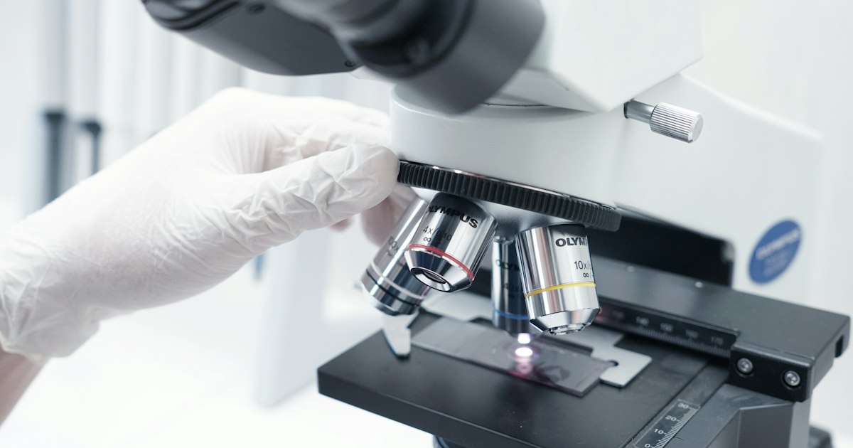
A drop of blood is spread thinly across a glass slide, stained (usually with Leishman or Giemsa stain in Indian labs), and examined under a microscope by a pathologist or trained technologist. Unlike an automated CBC, which counts cells, a smear lets an expert see them: their size, shape, colour, maturity and any abnormal cells or parasites.
How it differs from a CBC
| Feature | Complete Blood Count (CBC) | Peripheral Blood Smear |
|---|---|---|
| Done by | Automated analyser | Pathologist looking through a microscope |
| Gives you | Numbers: Hb, RBC, WBC, platelets, MCV, MCH, RDW | A written description of how cells look |
| Strength | Fast, precise counts | Spots abnormal shapes, immature cells, parasites |
| Can it miss things? | Yes, machines can be fooled by clumped platelets or unusual cells | Depends on the skill of the person reading it |
| Typical cost in India | ₹200–₹500 | ₹100–₹350 (often bundled with CBC) |
The two tests work best together. Many labs automatically do a smear when the analyser "flags" an abnormal CBC. Our CBC report guide explains the numbers; this article explains the words.
Why Doctors Order a Peripheral Smear
Your doctor may request a peripheral smear when:
- Haemoglobin is low and they need to know what kind of anaemia it is
- MCV is very high or very low, suggesting B12/folate deficiency or iron deficiency/thalassemia
- Platelets are low and they want to rule out a false reading from clumping
- White cell count is very high or very low, or the machine reports abnormal cells
- Fever raises suspicion of malaria, dengue or another infection
- There is unexplained jaundice, which may point to red cells breaking down (haemolysis)
- A child has a family history of thalassemia or sickle cell disease
- There are signs suggesting a blood cancer such as leukaemia, like persistent fever, bone pain, easy bruising or swollen glands
- Before or during treatment with medicines that can affect the bone marrow
How the Test Is Done: Preparation and Cost
Preparation
- No fasting is needed. You can eat and drink normally.
- Blood is drawn from a vein into a purple-top EDTA tube, the same tube used for a CBC. For malaria, a finger-prick sample may also be used.
- Tell the lab if you have had a blood transfusion in the last three months, as donor cells can change the picture.
- Mention any recent fever, and any medicines such as anti-epileptics, methotrexate, metformin or antibiotics.
Timing matters for accuracy
Smears are best made within 2 to 3 hours of collection. Blood that sits too long in an EDTA tube can develop artefacts, such as crenated (spiky) red cells or degenerated white cells, that a pathologist must learn to ignore. If you use home sample collection, it is reasonable to ask how quickly samples reach the lab.
Cost and turnaround
- Standalone peripheral smear: roughly ₹100 to ₹350 at chains such as SRL, Metropolis, Dr. Lal PathLabs and Thyrocare-partner labs, and much less at government hospitals.
- Often included free or at a small add-on cost with a CBC.
- Reports usually come the same day or next day, since a human must review the slide.
Decoding Your Peripheral Smear Report
A smear report usually has three parts: RBC morphology, WBC morphology and platelets, sometimes followed by an "impression" that summarises the likely cause.
Red blood cell (RBC) terms
| Term on report | What it means | Common causes in India |
|---|---|---|
| Normocytic normochromic | Normal size and colour | Normal, or anaemia from chronic disease, kidney disease or recent blood loss |
| Microcytic hypochromic | Small, pale cells | Iron deficiency, thalassemia trait, anaemia of chronic disease |
| Macrocytic / macro-ovalocytes | Large, often oval cells | Vitamin B12 or folate deficiency, alcohol, liver disease, hypothyroidism |
| Anisocytosis | Cells of varying size | Many anaemias, especially iron deficiency (matches a high RDW) |
| Poikilocytosis | Cells of varying shape | Many anaemias and bone marrow conditions |
| Pencil cells (elliptocytes) | Long, thin cells | Classic in iron deficiency |
| Target cells (codocytes) | Cells that look like a bullseye | Thalassemia, HbE trait, liver disease |
| Sickle cells | Crescent-shaped cells | Sickle cell disease, common in tribal belts of central and western India |
| Schistocytes | Fragmented cells | Red cells being torn in small vessels (e.g. TTP, HUS, severe infection); needs urgent attention |
| Spherocytes | Small, round, dense cells | Hereditary spherocytosis, autoimmune haemolysis |
| Basophilic stippling | Blue dots inside red cells | Thalassemia, lead poisoning |
| Nucleated RBCs | Immature red cells in blood | Severe anaemia, thalassemia major, marrow stress; normal in newborns |
| Polychromasia | Bluish young red cells | Marrow responding to blood loss or haemolysis, or to iron/B12 treatment |
| Rouleaux | Red cells stacked like coins | High protein levels, as in infection, inflammation or multiple myeloma |
White blood cell (WBC) terms
- Hypersegmented neutrophils: Neutrophils with six or more lobes, or many with five. This is one of the earliest signs of B12 or folate deficiency, sometimes appearing before MCV rises. See our folate guide.
- Toxic granulation / shift to left: Dark granules and younger neutrophils, typical of a bacterial infection or sepsis.
- Atypical (reactive) lymphocytes: Large, activated lymphocytes seen in viral infections such as dengue, infectious mononucleosis and hepatitis.
- Eosinophilia: More eosinophils than usual. In India, common causes are intestinal worms, allergies, asthma and tropical pulmonary eosinophilia.
- Blasts: Very immature white cells. Even a few blasts in the blood need prompt evaluation by a haematologist, as they can indicate leukaemia.
- Smudge cells: Fragile lymphocytes that break when the slide is made, often seen in chronic lymphocytic leukaemia in older adults.
Platelet terms
- Adequate on smear: The platelet count looks normal on the slide, even if the machine reported a low value.
- Platelet clumps seen: The machine likely undercounted platelets because they stuck together in the EDTA tube. This is called pseudothrombocytopenia and is harmless; a repeat in a citrate tube usually gives a normal count. Our low platelet count guide explains why this matters during dengue season.
- Giant platelets: Larger than usual platelets, seen when the marrow is releasing young platelets quickly (as in ITP or recovery from dengue) and in some inherited conditions.
Parasites
- Malaria parasites (MP): The smear can show Plasmodium vivax or falciparum and estimate how many cells are infected. A thick smear finds parasites, while a thin smear identifies the species. Rapid antigen tests are useful, but the smear remains the gold standard. Read more in our malaria test guide.
- Microfilariae: Larvae of filarial worms, best seen in a night blood sample taken between 10 pm and 2 am in endemic areas.
Common Smear Patterns Seen in Indian Patients
A single term rarely makes a diagnosis. Pathologists and doctors look at combinations of findings alongside your CBC and symptoms.
| Pattern on smear plus CBC | Most likely explanation | Usual next test |
|---|---|---|
| Microcytic hypochromic, pencil cells, high RDW | Iron deficiency anaemia | Serum ferritin, iron studies |
| Microcytic hypochromic, target cells, normal RDW, high RBC count | Thalassemia trait | HPLC / Hb electrophoresis |
| Macro-ovalocytes, hypersegmented neutrophils | B12 or folate deficiency | Serum B12, folate, homocysteine |
| Dimorphic picture (small and large cells together) | Mixed iron + B12 deficiency, very common in Indian vegetarians and pregnant women | Ferritin, B12, folate |
| Sickle cells, target cells | Sickle cell disease | HPLC, sickling test |
| Atypical lymphocytes, low platelets | Viral fever such as dengue | Dengue NS1/IgM, repeat CBC |
| Toxic granulation, high neutrophils | Bacterial infection | Cultures, CRP |
| Blasts, very high or low WBC | Possible acute leukaemia | Urgent haematology referral, bone marrow test |
| Schistocytes, low platelets | Microangiopathic process | Urgent medical review, LDH, kidney function |
The dimorphic picture deserves special mention for Indian readers. Because many Indians are vegetarian and many women enter pregnancy with low iron stores, it is common to have both iron and B12 deficiency at the same time. The two can cancel each other out on the MCV, which may look deceptively normal. Only the smear shows both populations of cells. See our guides on ferritin, thalassemia and sickle cell disease for the follow-up tests.
Limitations and When to Repeat the Test
What a smear cannot do
- It cannot measure iron or B12 levels directly; it only suggests a deficiency.
- It cannot confirm thalassemia trait; an HPLC test is needed.
- It depends heavily on the reader. Two pathologists may describe the same slide slightly differently.
- It can miss low-level malaria parasites, which is why a negative smear in a feverish patient is often repeated.
When a repeat smear helps
- Fever with a negative malaria smear: Repeat every 12 to 24 hours if symptoms continue, as advised by your doctor.
- After starting iron or B12: A smear can show young red cells (polychromasia) appearing within one to two weeks, an early sign that treatment is working.
- Low platelets with clumps: A repeat CBC in a citrate tube confirms the true count.
- Abnormal cells seen: Your haematologist may ask for a fresh sample, a repeat smear, or flow cytometry before deciding on a bone marrow test.
Always discuss your smear report with your doctor. Some terms, like occasional target cells or mild anisocytosis, are minor findings, while others, like blasts or schistocytes, need same-day attention.
Keeping Smear Reports With Your Blood Count History
The peripheral smear is a descriptive report, which makes it easy to lose track of. Unlike a haemoglobin value, you cannot easily plot "microcytic hypochromic" on a graph. But the descriptions change meaningfully over time. A child with thalassemia trait, a vegetarian adult on B12 injections, or an elderly parent being watched for a blood disorder will have a series of smears that tell a story.
When you upload your reports to MedicalVault, the smear report stays attached to the CBC from the same day, so your doctor sees the numbers and the morphology together. MedicalVault's trend analysis charts your haemoglobin, MCV and platelets over months, so you can see whether iron or B12 treatment is actually working. And if you are managing a parent's blood tests from another city, the family sharing feature lets you open their latest report before their next appointment.
Key Takeaways
- A peripheral blood smear test is a microscope review of your blood that describes the size, shape and maturity of red cells, white cells and platelets, and can detect parasites like malaria.
- It costs only ₹100–₹350 in India, needs no fasting, and is best done together with a CBC.
- Microcytic hypochromic cells with pencil forms suggest iron deficiency; macro-ovalocytes with hypersegmented neutrophils suggest B12 or folate deficiency; target cells point toward thalassemia or liver disease.
- A dimorphic picture (iron plus B12 deficiency together) is common in Indian vegetarians and pregnant women and can hide behind a normal MCV.
- Platelet clumps can make a CBC falsely show low platelets; the smear catches this.
- Blasts or schistocytes on a smear need urgent medical attention. Always review your report with your doctor.
- Keep every smear and CBC together in MedicalVault so your doctor can see both the numbers and the descriptions over time.