Your doctor writes "CT abdomen with contrast" on a prescription pad, and within the hour you are standing at a diagnostic centre counter being quoted ₹6,500, asked whether you want it "with report same day", and handed a consent form full of words like nephrogenic and extravasation. Two days later you collect a film and a typed page that ends with the sentence every Indian patient has misread at least once: "Correlate clinically."
India runs an enormous volume of scans — there are well over 40,000 imaging centres nationwide, and prices for the exact same MRI can differ fourfold between a Mumbai corporate hospital and a standalone centre in Lucknow. Yet almost nobody explains which scan answers which question, how much radiation you are actually getting, when contrast is risky, or what the phrases in your report mean. This guide covers all of it: X-ray, ultrasound, CT and MRI — what each one sees, what each costs in India, how to read the report, and how to avoid paying for scans you did not need.
Four Scans, Four Completely Different Jobs
The single most useful thing to understand is that these machines do not compete — they see different things.
| Scan | How It Works | Best For | Blind Spots |
|---|---|---|---|
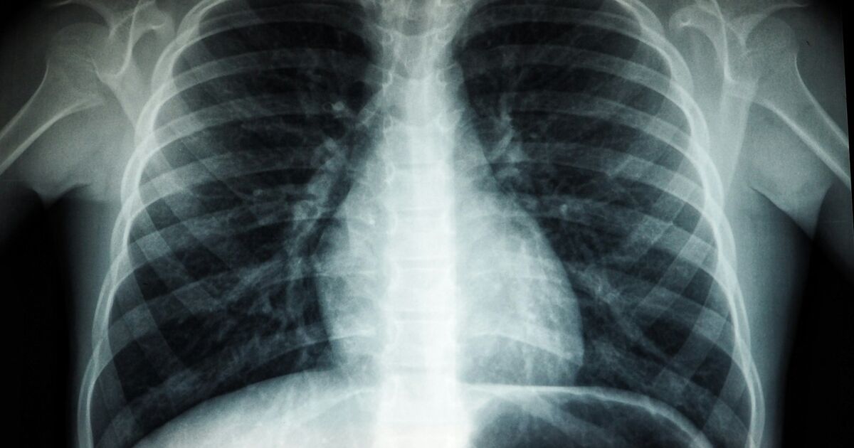
| X-ray | Radiation passes through the body onto a detector | Bones, fractures, chest/lungs, dental, blockages in the gut | Poor at soft tissue, ligaments, early cartilage damage |
| Ultrasound (USG) | Sound waves, no radiation at all | Abdomen, liver, kidneys, gallbladder, pregnancy, thyroid, neck lumps, blood flow | Cannot see through bone or gas — poor for lungs, brain, deep bowel |
| CT scan | Many X-rays combined into cross-sections | Emergencies, head injury, stroke, chest, abdomen, kidney stones, lungs, bone detail | Uses the most radiation; less detail in soft tissue than MRI |
| MRI | Strong magnet plus radio waves, no radiation | Brain, spinal cord, discs, ligaments, joints, soft tissue tumours | Slow, noisy, expensive; unsafe with some metal implants |
A few consequences of this that save money and worry:
- Knee pain with a suspected ligament tear needs an MRI, not an X-ray. An X-ray of the knee will look "normal" while a fully torn ACL sits there undetected. X-ray is for bone.
- A suspected kidney stone usually needs a non-contrast CT (CT KUB) — it picks up stones an ultrasound will miss, particularly in the ureter.
- Abdominal pain in a child, or anything in pregnancy, starts with ultrasound because there is no radiation.
- Stroke needs an urgent CT head first — not to show the stroke, but to rule out bleeding before clot-busting treatment can be given. Read our stroke guide for why the first hour matters.
Where Do PET, Mammography and DEXA Fit?
| Scan | What It Is For |
|---|---|
| PET-CT | Cancer staging and response assessment; combines metabolic activity with anatomy |
| Mammography | Breast cancer screening and evaluation of lumps (see our breast cancer guide) |
| DEXA | Bone mineral density for osteoporosis |
| Doppler | An ultrasound add-on showing blood flow — used for DVT, varicose veins, carotids |
| CT angiography / coronary CT | Blood vessels, including the coronary calcium score |
Radiation: The Honest Numbers
This is where Indian patients get either falsely terrified or dangerously casual. Radiation dose is measured in millisieverts (mSv). Everyone in India already absorbs roughly 2–3 mSv a year from natural background sources — soil, cosmic rays, food.
| Examination | Approximate Dose (mSv) | Roughly Equivalent To |
|---|---|---|
| Ultrasound | 0 | Nothing |
| MRI | 0 | Nothing |
| Dental X-ray | 0.005 | Under a day of background radiation |
| Chest X-ray | 0.1 | About 10 days of background |
| Mammogram | 0.4 | About 7 weeks |
| X-ray abdomen | 0.7 | About 4 months |
| CT head | 2 | About 8 months |
| CT chest | 6–7 | About 2 years |
| CT abdomen and pelvis | 8–10 | About 3 years |
| PET-CT | 15–25 | 5–8 years |
What to take from this table:
- One CT scan when it is medically indicated is worth it. The information gained almost always outweighs a theoretical long-term risk.
- Repeated CTs are a genuine concern, particularly in children, teenagers and young adults, whose tissues are more radiosensitive and who have more years ahead for a risk to express itself.
- There is no radiation in ultrasound or MRI at all. If a family member is anxious, this is the reassurance to give them.
- Ask the question that actually helps: "Doctor, will this scan change what we do next?" If the answer is no, the scan probably is not needed.
In India, every centre using X-ray or CT equipment must be registered with the Atomic Energy Regulatory Board (AERB), which sets equipment and shielding standards. A centre that cannot show AERB registration is one to walk away from. MRI and ultrasound do not fall under AERB since they use no ionising radiation — though ultrasound centres must display PCPNDT Act registration, and are legally barred from disclosing the sex of a foetus.
Contrast: When It Helps and When It Is Risky
"With contrast" means a dye is injected (or swallowed) to make blood vessels, inflammation and tumours stand out. It roughly doubles the cost and adds a layer of risk that deserves a real conversation.
| Type | Used In | Main Concerns |
|---|---|---|
| Iodinated contrast | CT | Allergic reaction; can stress the kidneys (contrast-induced nephropathy) |
| Gadolinium | MRI | Rarely allergic; unsafe with severe kidney impairment (eGFR under 30) |
| Barium | X-ray studies of gut | Constipation; not used if a perforation is suspected |
| Microbubble | Contrast ultrasound | Very low risk overall |
What Your Centre Must Know Before a Contrast Study
Tell the radiology staff, in advance and without waiting to be asked:
- Your kidney function. Most centres require a recent serum creatinine or eGFR before contrast. If you have not had one, our kidney function test guide explains what it measures.
- Any previous reaction to contrast, iodine or seafood.
- Asthma or significant allergies — these raise reaction risk modestly.
- Metformin, sold in India as Glycomet, Glyciphage, Obimet and dozens of combination brands. Doctors often pause it around an iodinated-contrast CT in people with reduced kidney function.
- Pregnancy, or any chance of it.
- Breastfeeding — generally safe to continue, but confirm with your radiologist.
Most reactions are mild: a warm flush, a metallic taste, mild nausea. These are expected, not emergencies. Serious reactions are rare, and every accredited centre keeps emergency drugs and trained staff on hand.
MRI-Specific Safety
MRI is a very powerful magnet that is always on, even between scans. Declare all of the following:
- Pacemaker or implanted defibrillator — many modern ones are MRI-conditional, but this needs planning, not a same-day decision
- Cochlear implant, aneurysm clips, certain older heart valves
- Metal fragments in the eye (a real risk for welders and factory workers — an orbital X-ray may be done first)
- Orthopaedic plates, screws, joint replacements, stents — usually safe, but declare them anyway
- Claustrophobia — ask about open or wide-bore machines, or mild sedation; the tunnel is narrow and the scan is loud for 20–45 minutes
Reading Your Radiology Report Without Panicking
Reports follow a standard structure, and knowing it defuses most of the fear.
| Section | What It Contains |
|---|---|
| Clinical history / indication | Why the scan was ordered |
| Technique | Machine, sequences, contrast used |
| Findings | Everything the radiologist observed, organ by organ |
| Impression / Conclusion | The radiologist's actual summary — read this first |
Most patients read the findings section, panic at an unfamiliar word, and never reach the impression. Reverse the order.
Common Report Phrases, Translated
| Phrase | What It Usually Means |
|---|---|
| "No significant abnormality detected" | Normal for the question asked |
| "Correlate clinically" | "This finding only matters alongside your symptoms and examination" — not a warning |
| "Incidental finding" | Something spotted by accident, unrelated to why you came |
| "Fatty infiltration of liver" / Grade I–III | Fatty liver — very common in India; see our fatty liver guide |
| "Simple cyst" | Fluid-filled sac; usually harmless, often needs nothing |
| "Lesion" / "SOL" | A neutral word for any abnormal area — not a synonym for cancer |
| "Calcified granuloma" | An old healed infection, very often past TB — extremely common in Indians, needs no treatment |
| "Degenerative changes" / "spondylosis" | Age-related wear; present in most adults over 40, often painless |
| "Disc bulge" | Common and frequently unrelated to back pain; see our low back pain guide |
| "Suggestive of" / "cannot be excluded" | Genuine uncertainty — further tests or follow-up imaging will decide |
| "Prominent" / "borderline" | At the edge of normal; usually watched, not treated |
Two habits worth building. First, never diagnose yourself from a radiology report. Radiologists describe shadows; doctors diagnose patients. A report saying "cannot rule out malignancy" is a call for the next test, not a verdict. Second, always take the images, not only the report, to your consultation — on CD, film or a link. A second radiologist or your treating doctor may read the same images differently, which is a normal and healthy part of medicine.
Scan reports are also the single most commonly lost document in Indian households. CDs stop being readable, films fade and crack, and the one-page report goes missing exactly when a new doctor asks "was there a nodule on the old CT?" When you upload your scan reports to MedicalVault, the report text is stored and searchable, so a 2023 chest CT finding can be pulled up in an OPD queue in seconds rather than reconstructed from memory.
What Scans Cost in India — and How to Pay Less
Prices vary enormously by city, machine and whether a hospital is attached.
| Scan | Typical Range (₹) |
|---|---|
| Chest X-ray | 200 – 600 |
| X-ray, other regions | 300 – 900 |
| Ultrasound abdomen/pelvis | 800 – 2,500 |
| Doppler study | 1,500 – 4,000 |
| CT head (plain) | 1,500 – 4,000 |
| CT chest / abdomen (plain) | 2,500 – 7,000 |
| CT with contrast | 4,000 – 12,000 |
| MRI, single region (plain) | 3,000 – 12,000 |
| MRI with contrast | 6,000 – 20,000 |
| PET-CT (whole body) | 18,000 – 35,000 |
| Mammography | 1,200 – 3,500 |
Practical ways to reduce what you pay:
- Standalone diagnostic centres are usually cheaper than hospital radiology departments for the same scan on a comparable machine.
- Tier-2 cities cost substantially less. The same MRI that is ₹12,000 in south Mumbai can be ₹5,000–6,000 in Nagpur or Lucknow.
- Government medical colleges and district hospitals offer imaging free or at token rates, with a waiting list.
- Ayushman Bharat PM-JAY covers diagnostics bundled within an admitted treatment package — read our PM-JAY guide for eligibility.
- Outpatient scans are rarely covered by private health insurance unless you have an OPD rider; scans done during an admission usually are. Our health insurance claims guide covers the documentation to keep.
- Ask whether a plain scan will do. Contrast roughly doubles the bill and is not always needed.
- Do not repeat a scan simply because a new doctor has not seen it. Carry the previous images. This alone saves Indian families thousands of rupees a year.
Questions Worth Asking at the Counter
- Is the centre AERB registered, and is a qualified radiologist reporting it?
- Is a radiologist's typed report included, or only films?
- Will I get the images digitally, or only on a CD?
- What is the MRI machine strength — 1.5 Tesla or 3 Tesla? (Higher is not always needed, but matters for brain and joint work.)
- How long until the report, and can it be sent by email or WhatsApp?
Key Takeaways
- X-ray sees bone, ultrasound sees soft organs without radiation, CT is fast and detailed, MRI is best for brain, spine, joints and soft tissue. Choosing the right one first avoids paying twice.
- Ultrasound and MRI involve zero radiation. A CT abdomen carries roughly 8–10 mSv — worth it when indicated, worth questioning when repeated.
- Always ask your doctor: "Will this scan change what we do next?"
- Declare kidney function, allergies, metformin, pregnancy and any implanted metal before a contrast or MRI study.
- Read the Impression first, not the findings. "Correlate clinically" is routine phrasing, not a warning, and "lesion" does not mean cancer.
- The same scan can cost four times more in one city than another; standalone centres and tier-2 cities are consistently cheaper.
- Carry your old images to every consultation — and keep them permanently with MedicalVault's report storage and trend analysis, so a comparison scan is always one search away instead of one repeat scan away.
Always discuss which scan you need, and what the report means, with your treating doctor — a radiology report is one piece of evidence, never a diagnosis on its own. To understand how artificial intelligence is changing scan reporting in India, read our AI in radiology and pathology guide, and see the features page for how MedicalVault keeps every report your family collects in one searchable place.