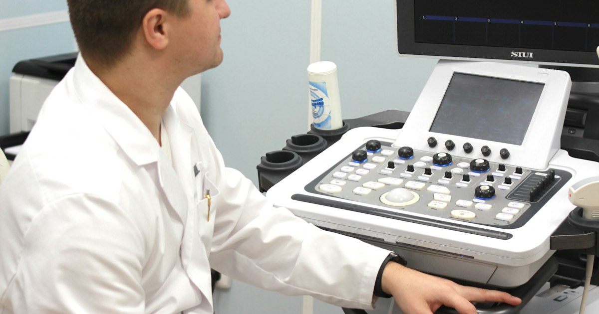
You went to the doctor with a vague stomach ache, and you walked out with a slip that said "USG whole abdomen". Two hours later, the report is in your hand: "Liver is enlarged with diffusely increased echogenicity, suggestive of Grade I fatty liver. Gallbladder shows a 6 mm calculus. Right kidney shows a 3 mm concretion." Your follow-up appointment is three days away, and in the meantime, every word on that page has been typed into a search engine.
The ultrasound whole abdomen, or USG abdomen, is one of the most commonly ordered scans in India. It is cheap, painless, uses no radiation and is available from district hospitals to neighbourhood diagnostic centres. It is also the scan that most often surprises people with findings they never had symptoms for, like fatty liver, gallstones, kidney stones or a simple cyst. With an estimated 38% of Indian adults having fatty liver according to a 2022 meta-analysis, a large share of USG reports will carry at least one finding. This guide explains what the scan covers, how to prepare, normal sizes, what common report terms mean, which findings are usually harmless and which need prompt attention.
What Is an Ultrasound Whole Abdomen?
An ultrasound uses high-frequency sound waves sent by a handheld probe (transducer). The waves bounce off organs and are converted into a real-time image. Because it uses no X-rays, it is safe in pregnancy and for children, and it can be repeated as often as needed.
A USG whole abdomen usually includes both the upper abdomen and the pelvis:
| Region | Organs examined |
|---|---|
| Upper abdomen | Liver, gallbladder, bile ducts (CBD), pancreas, spleen, portal vein, aorta |
| Kidneys and retroperitoneum | Both kidneys, ureters (if dilated), adrenal region, lymph nodes |
| Pelvis | Urinary bladder; prostate in men; uterus and ovaries in women |
| General | Free fluid (ascites), bowel loops, hernias in some cases |
Some doctors order a narrower study, such as USG upper abdomen (liver, gallbladder, pancreas, spleen) or USG KUB (kidneys, ureters and bladder, with prostate in men), when they are looking for a specific problem.
Why doctors order it
- Abdominal pain, especially in the upper right abdomen (gallbladder, liver) or flank (kidneys)
- Abnormal blood tests, such as raised liver enzymes in a liver function test or raised creatinine
- Urinary symptoms: burning, blood in urine, frequent urination at night, difficulty passing urine
- Jaundice, unexplained weight loss, vomiting or a lump in the abdomen
- Routine health check-ups, where it is often included in packages for adults above 40
- Follow-up of known fatty liver, gallstones, kidney stones, cysts or fibroids
Cost of USG abdomen in India
In private diagnostic centres, a USG whole abdomen typically costs ₹800 to ₹2,000, depending on the city and centre. Government hospitals offer it free or at very low cost, though waiting lists can be long. It is covered under schemes such as CGHS and is often part of preventive health packages.
How to Prepare for a USG Abdomen
Good preparation makes a real difference to how clearly the radiologist can see your organs.
| Preparation | Why it matters |
|---|---|
| Fast for 6 to 8 hours (usually overnight) | The gallbladder contracts after food and cannot be assessed properly. Food also increases bowel gas, which blocks the view of the pancreas |
| Drink 4 to 6 glasses of water 45 to 60 minutes before and do not pass urine | A full bladder pushes gas-filled bowel aside and allows the bladder, prostate, uterus and ovaries to be seen |
| Avoid fizzy drinks, chewing gum and smoking before the scan | These increase swallowed air and bowel gas |
| Continue regular medicines with sips of water, unless told otherwise | People with diabetes should ask their doctor about adjusting medicines while fasting |
| Carry old reports and scans | The radiologist can compare sizes of stones, cysts or fibroids |
| Wear loose clothing | You may be asked to lower the waistband or change into a gown |
If you have diabetes, book an early morning slot so that fasting does not cause low blood sugar. Children and elderly people may be given shorter fasting times.
The scan itself takes about 15 to 30 minutes. Gel is applied on your abdomen, and you may be asked to take a deep breath and hold it, or turn on your side. You may be asked to empty your bladder at the end so that the post-void residual (PVR) urine can be measured.
A note for women
Under India's PCPNDT Act, ultrasound centres must be registered, and determining or revealing the sex of a foetus is a criminal offence. Pregnant women will be asked to fill a Form F. This is routine and protects against misuse. Pregnancy scans are different from a whole abdomen scan; our antenatal care and pregnancy tests guide covers pregnancy scans and tests.
Normal Sizes in a USG Abdomen Report
Every report describes the size, shape, outline and echotexture (the internal pattern of the organ). The values below are typical adult ranges; exact cut-offs vary with age, body size and the radiologist's reference.
| Organ | Typical normal value in adults | What is reported if abnormal |
|---|---|---|
| Liver | Up to about 15 cm (craniocaudal span); smooth outline, uniform echotexture | Hepatomegaly, fatty infiltration, coarse echotexture, nodular surface |
| Gallbladder wall | Less than 3 mm when fasting | Wall thickening, calculi (stones), sludge, polyp |
| Common bile duct (CBD) | Under 6 mm (up to about 1 mm more per decade after 60; wider after gallbladder removal) | Dilated CBD, choledocholithiasis (stone in the duct) |
| Portal vein | Up to about 13 mm | Dilated portal vein, a sign of portal hypertension |
| Spleen | Up to about 12 to 13 cm | Splenomegaly |
| Kidneys | About 9 to 12 cm long, with clear corticomedullary differentiation | Calculus, hydronephrosis, cyst, raised echogenicity, small kidneys |
| Urinary bladder | Smooth, thin wall when full | Wall thickening, calculus, high post-void residual |
| Post-void residual (PVR) | Under about 50 ml (up to 100 ml often accepted in older adults) | Incomplete emptying |
| Prostate | About 20 to 25 ml (cc) in younger adult men | Prostatomegaly (enlarged prostate) |
| Uterus | Size varies with age and childbirth history | Bulky uterus, fibroids, adenomyosis, thickened endometrium |
| Ovaries | Size varies with age and cycle | Ovarian cyst, polycystic morphology |
A line such as "No obvious abnormality detected" or "Normal study" is the result most people hope for. "Pancreas obscured by bowel gas" simply means the radiologist could not see it fully, which is common and not a finding in itself. If there is a specific concern about the pancreas, your doctor may repeat the scan after better preparation or order a CT.
Common USG Report Terms Explained
Ultrasound reports use specialised language. Here is what common phrases usually mean.
| Report term | Plain meaning |
|---|---|
| Echogenicity / echotexture | How bright the organ looks and how uniform its internal pattern is |
| Hyperechoic / echogenic | Brighter than normal, as seen in fatty liver, stones or some lesions |
| Hypoechoic / anechoic | Darker than normal; anechoic (black) usually means fluid, as in a simple cyst |
| Posterior acoustic shadowing | A dark shadow behind a structure, typical of stones |
| Hepatomegaly / splenomegaly | Enlarged liver / enlarged spleen |
| Cholelithiasis | Gallstones |
| Sludge | Thickened bile in the gallbladder |
| Nephrolithiasis / renal calculus | Kidney stone |
| Concretion / echogenic focus | A tiny bright spot in the kidney, often a very small stone or calcification |
| Hydronephrosis | Swelling of the kidney's collecting system, usually due to blockage |
| CMD maintained | Normal corticomedullary differentiation, a reassuring sign for kidney health |
| Ascites / free fluid | Fluid in the abdomen |
| Prostatomegaly | Enlarged prostate |
| Bulky uterus | A uterus larger than average, often due to childbirth, fibroids or adenomyosis |
Fatty liver grades
Fatty liver is by far the most common finding on Indian USG reports. Radiologists grade it by how bright the liver looks and how well deeper structures can be seen:
| Grade | Ultrasound appearance | What it usually means |
|---|---|---|
| Grade I (mild) | Liver slightly brighter; vessels and diaphragm clearly seen | Early fat build-up, usually reversible with lifestyle change |
| Grade II (moderate) | Liver brighter; portal vein walls and diaphragm less clearly seen | More fat; needs evaluation of liver enzymes and metabolic risk |
| Grade III (severe) | Markedly bright; diaphragm and back of liver poorly seen | Significant fat; needs medical review |
An important point: ultrasound grades fat, not damage. A Grade I report does not rule out scarring (fibrosis), and a Grade III report does not mean cirrhosis. Doctors use blood tests, the FIB-4 score and FibroScan to assess scarring. Our fatty liver guide explains the next steps.
Which Findings Need Attention?
Many USG findings are incidental and harmless. Others need prompt action. This is general guidance; always discuss your report with your doctor.
Usually not serious (but discuss at your next visit)
- Grade I fatty liver with normal liver enzymes: a warning sign for diabetes and heart disease, best handled with diet, exercise and weight loss
- Simple renal cyst: a thin-walled, fluid-filled sac in the kidney, very common after 50 and usually harmless
- Small hepatic haemangioma: a benign cluster of blood vessels in the liver, usually needing no treatment
- Tiny renal concretions (under 4 to 5 mm) without hydronephrosis, which may pass on their own
- Mild prostatomegaly in older men without significant urinary symptoms
- Small physiological ovarian cysts and a small amount of free fluid in the pelvis around ovulation in women of reproductive age
- Gallbladder polyps under 10 mm, which usually need periodic follow-up scans
Needs a doctor's review soon
- Gallstones, especially with pain after meals; see our gallstones guide
- Kidney stones larger than 5 mm, or any stone with hydronephrosis; our kidney stones guide covers treatment options
- Grade II or III fatty liver, coarse echotexture, or an irregular liver surface
- Splenomegaly or a dilated portal vein
- Enlarged prostate with high PVR (over 100 ml), or difficulty passing urine; your doctor may also check a PSA test
- Fibroids, complex ovarian cysts or polycystic ovaries; see our PCOS guide
- Raised kidney echogenicity or small kidneys, which may indicate chronic kidney disease
Seek urgent care
- Dilated CBD with jaundice, fever or severe pain
- Gallbladder wall thickening with fluid around it and pain, which may indicate acute cholecystitis
- Inflamed appendix or free fluid with severe pain
- Significant ascites
- Any solid mass or lesion in the liver, kidney, pancreas or ovary that the report recommends evaluating with CT or MRI
Limitations of ultrasound
Ultrasound is excellent for many problems, but it is operator-dependent, meaning results depend on the skill of the person scanning and the quality of the machine. Bowel gas, obesity and poor preparation can hide organs. It is not a good test for the bowel itself, and small lesions may be missed. When needed, doctors follow up with a CT scan or MRI; our X-ray, CT and MRI guide explains how these compare.
Comparing Your USG Reports Over Time
Ultrasound measurements are most useful when compared with previous scans. Is the gallstone the same size as last year? Has the fatty liver improved from Grade II to Grade I after weight loss? Has the prostate grown, and is the PVR rising? Is the fibroid getting bigger? A single report shows a snapshot; a series of reports shows the direction.
Yet most families keep USG reports as thermal-paper printouts that fade within months, or as photographs scattered across phones. When you upload your reports to MedicalVault, your USG findings sit alongside your blood tests, so your doctor can see the full picture: your liver ultrasound next to your liver enzymes, or your kidney scan next to your creatinine. MedicalVault's trend analysis shows how values such as ALT, HbA1c and creatinine change as you work on fatty liver or kidney health. With the family sharing feature, you can keep a parent's prostate or kidney scans organised and share them with a specialist before an appointment.
Practical tips for your next scan
- Ask for the report and key images, either on paper or digitally
- Get repeat scans at the same centre when possible, to reduce variation in measurements
- Note the exact size of stones, cysts, fibroids or prostate volume, so you can compare
- Do not change medicines or start treatments based on the report alone; consult your doctor
Key Takeaways
- A USG whole abdomen checks the liver, gallbladder, bile ducts, pancreas, spleen, kidneys, bladder and pelvic organs, without radiation, and typically costs ₹800 to ₹2,000 in private centres.
- Fast for 6 to 8 hours and come with a full bladder for the clearest images; avoid fizzy drinks and chewing gum before the scan.
- Typical adult values include a liver up to about 15 cm, spleen up to 12 to 13 cm, kidneys 9 to 12 cm, CBD under 6 mm and a post-void residual under 50 ml.
- Fatty liver is the most common finding in India; ultrasound grades fat but not scarring, so FibroScan and blood tests are needed to assess damage.
- Simple renal cysts, small haemangiomas and small physiological ovarian cysts are usually harmless, while gallstones with pain, stones with hydronephrosis, a dilated CBD or any solid mass need a doctor's review.
- Ultrasound is operator-dependent and may miss small lesions; CT or MRI may be advised for unclear findings.
- Keep every USG report in MedicalVault so you and your doctor can compare stone sizes, fatty liver grades and prostate volume over the years.