Imagine losing your eyesight — not because of an accident, but because of diabetes you thought was under control. This is the silent reality for hundreds of thousands of Indians every year. Diabetic retinopathy is the leading cause of preventable blindness in working-age adults in India, and it has one defining, terrifying feature: it causes no symptoms until it's already quite advanced.
India is home to over 10 crore people with diabetes — the second highest number in the world. The SMART India study published in The Lancet Global Health found that 12.5% of people with diabetes in India have diabetic retinopathy, and 4% have the vision-threatening form. That translates to roughly 1.2 crore Indians with some degree of diabetic eye damage — most of whom don't know it. The 2023 joint guidelines from RSSDI (Research Society for the Study of Diabetes in India) and VRSI (Vitreoretinal Society of India) made it explicit: every person with diabetes in India needs annual eye screening, regardless of how well-controlled their sugar is.
If you or a family member has diabetes, this article could protect their sight.
What is Diabetic Retinopathy?
The retina is the light-sensitive layer at the back of your eye — think of it as the "film" in a camera. It's nourished by a dense network of tiny blood vessels. Chronically high blood sugar damages these vessels in four ways:
- They leak: Fluid and blood ooze into the retina, causing swelling (oedema) and blurred vision
- They block: Vessels become occluded, cutting off oxygen to parts of the retina
- They break: New, fragile blood vessels grow to compensate (neovascularisation) — but these new vessels bleed easily, causing sudden vision loss
- The macula swells: The central retina (macula), responsible for sharp vision, fills with fluid — this is Diabetic Macular Oedema (DME) and is the most common cause of vision loss from diabetes
Diabetic retinopathy is classified by severity:
| Stage | Name | What's Happening | Vision at Risk? |
|---|---|---|---|
| Stage 1 | Mild NPDR | Tiny microaneurysms (bulges) in vessels | No |
| Stage 2 | Moderate NPDR | More vessel damage, haemorrhages, exudates | No |
| Stage 3 | Severe NPDR | Large areas of retina losing blood supply | Yes — progresses rapidly |
| Stage 4 | PDR (Proliferative) | New blood vessels forming on the retina | Imminent — emergency treatment needed |
| + | DME | Macular swelling at any stage | Yes — immediate treatment |
NPDR = Non-Proliferative Diabetic Retinopathy; PDR = Proliferative Diabetic Retinopathy
The key insight: Stages 1 and 2 are asymptomatic. You can have moderate NPDR — significant retinal damage — and still see perfectly. By the time vision blurs, you may already have progressed to the vision-threatening stage.
Who is Most at Risk? The Indian Picture
The risk of diabetic retinopathy increases dramatically with:
- Duration of diabetes: After 15 years of Type 2 diabetes, nearly 1 in 3 Indians has retinopathy. After 20+ years, prevalence can exceed 50%.
- Poor glycaemic control: Every 1% rise in HbA1c increases the risk of retinopathy progression by ~30%. Keeping HbA1c below 7% is the single most important protective factor.
- High blood pressure: Uncontrolled hypertension dramatically accelerates vessel damage in the retina
- High cholesterol: Elevated LDL accelerates exudate formation and macular damage
- Kidney disease: If your kidneys are affected by diabetes (diabetic nephropathy), your eyes almost certainly are too — the same microvascular damage mechanism affects both organs
- Type 1 diabetes: Near-universal retinopathy after 20 years; screening should begin 5 years after diagnosis
Indian-specific risk factors: Indians tend to develop Type 2 diabetes at a younger age and at lower BMI than Western populations — meaning longer lifetime exposure to high blood sugar. Indians also have poorer average glycaemic control than many developed nations, with a significant proportion unaware they even have diabetes. This combination makes Indian diabetics more vulnerable to retinopathy complications at relatively younger ages.
Symptoms: Why Diabetic Retinopathy is Called the "Silent Thief of Sight"
In the early stages — which can last years — diabetic retinopathy causes no symptoms at all. The first symptoms typically appear only in advanced disease:
- Blurring or fluctuating vision (often the first sign of diabetic macular oedema)
- Floaters: Spots or strings floating in your field of vision — often caused by small bleeds into the vitreous gel
- Flashes of light: Can indicate traction on the retina
- Dark or empty areas in your central vision
- Sudden, complete, or near-complete vision loss: This is a vitreous haemorrhage — a medical emergency
Critical point: By the time these symptoms appear, significant damage has already occurred. Treatment can still preserve remaining vision, but it cannot fully reverse damage. This is why proactive screening before symptoms develop is non-negotiable.
How Diabetic Retinopathy is Diagnosed
The Fundus Examination: What Your Eye Doctor Does
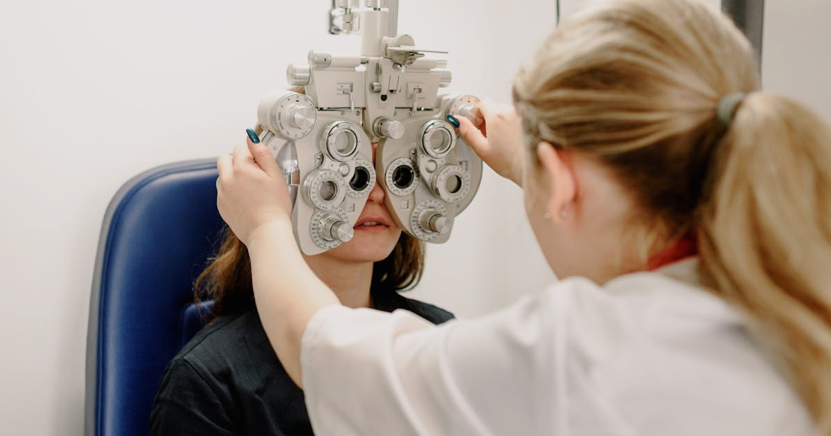
The definitive diagnostic test for diabetic retinopathy is a fundus examination — a direct look at the retina using specialised equipment. Your ophthalmologist uses one of these methods:
1. Fundus Photography (Retinal Photography)
A fundus camera takes high-resolution photographs of your retina. This is the preferred screening method in India because:
- It creates a visual record you can compare year to year
- Results can be reviewed remotely (teleophthalmology)
- AI-powered analysis software is increasingly available to assist screening
- No pupil dilation required with non-mydriatic cameras
Where to get it in India: Most major eye hospitals (LV Prasad Eye Institute, Aravind Eye Hospitals, Sankara Nethralaya, Shroff's Eye Centre), private ophthalmology clinics, and some diabetology clinics. Cost: ₹300–₹2,000 depending on location.
2. Dilated Fundus Examination (Indirect Ophthalmoscopy)
Your ophthalmologist dilates your pupils with eye drops and examines the retina directly. This allows examination of the peripheral retina that fundus cameras sometimes miss. Dilation takes 20–30 minutes to take effect; avoid driving afterwards.
3. OCT (Optical Coherence Tomography): The Gold Standard for Macular Assessment
OCT uses light waves to create a cross-sectional scan of the macula, similar to an ultrasound but using light instead of sound. It is the most sensitive test for detecting Diabetic Macular Oedema (DME) — even before it affects vision.
| OCT Finding | What It Means |
|---|---|
| Normal macular thickness (<250 μm) | No macular oedema |
| Increased thickness with fluid pockets | Centre-Involving DME — treatment warranted |
| Hard exudates near the macula | Risk of DME progression |
| Vitreomacular traction | May need vitreoretinal surgery |
OCT cost in India: ₹500–₹2,500 at most eye hospitals.
4. Fundus Fluorescein Angiography (FFA)
A dye (fluorescein) is injected intravenously and sequential photographs track it moving through retinal blood vessels. FFA is the best test to identify:
- Areas of retinal ischaemia (poor blood supply)
- Neovascularisation (new vessel growth in PDR)
- Microaneurysm leakage
- Guide for laser photocoagulation treatment
FFA is used when treatment planning is needed, not for routine screening. Cost: ₹1,500–₹5,000.
The Screening Schedule Every Diabetic in India Should Follow
The 2023 RSSDI–VRSI joint guidelines recommend:
| Type of Diabetes | When to Start Screening | Frequency |
|---|---|---|
| Type 2 diabetes | At diagnosis | Annually if no DR; every 6 months if early DR present |
| Type 1 diabetes | 5 years after diagnosis | Annually |
| Gestational diabetes | At diagnosis of GDM | Every trimester during pregnancy; 1 year postpartum |
| Known NPDR (mild) | Already under care | Annually |
| Known moderate/severe NPDR | Already under care | Every 6 months |
| Active PDR or DME | Already under care | Every 3 months or as directed |
Key message: If you were diagnosed with Type 2 diabetes today, you should have a dilated eye exam or fundus photograph today — because many people have had silent diabetes for years before diagnosis, and retinopathy may already be present.
Treatment Options: From Laser to Injections
Fortunately, if caught early, diabetic retinopathy is highly treatable. The treatment options in India include:
1. Optimise Blood Sugar, BP, and Cholesterol (Foundation of Treatment)
No eye treatment is effective if the underlying metabolic problems are not controlled. Achieving HbA1c <7%, blood pressure <130/80 mmHg, and LDL <100 mg/dL significantly slows or halts retinopathy progression. Track all these regularly using MedicalVault's trend analysis to spot deterioration before your next clinic visit.
2. Laser Photocoagulation (Panretinal Photocoagulation — PRP)
Laser photocoagulation destroys peripheral retinal tissue that is poorly oxygenated, reducing the stimulus for new, dangerous blood vessel growth. It is the standard treatment for Proliferative Diabetic Retinopathy.
- Type: Focal laser (for DME) or Panretinal Photocoagulation (for PDR)
- Sessions needed: 1–3 sessions typically, each lasting 20–30 minutes
- Does it hurt?: Mild discomfort; done under topical anaesthetic eye drops
- Side effects: Mild reduction in peripheral/night vision; transient visual blur
- Cost in India:
- Government hospitals (AIIMS, LV Prasad): ₹500–₹2,000 per session
- Private eye hospitals: ₹8,000–₹25,000 per session
- Approximate total (all sessions): ₹15,000–₹50,000
3. Anti-VEGF Injections: The Revolution in Macular Treatment
VEGF (Vascular Endothelial Growth Factor) is the chemical trigger that causes leaky blood vessels and new vessel growth in diabetic retinopathy. Anti-VEGF drugs block this trigger.
Anti-VEGF injections into the vitreous gel of the eye are now the first-line treatment for centre-involving Diabetic Macular Oedema and are used alongside laser for PDR.
| Drug | Indian Brand Names | Cost per Injection |
|---|---|---|
| Ranibizumab | Accentrix (Novartis), Razumab (Intas) | ₹20,000–₹55,000 |
| Bevacizumab | Avastin (off-label use) | ₹3,000–₹8,000 |
| Aflibercept | Eylea (Bayer) | ₹35,000–₹60,000 |
| Faricimab | Vabysmo (Roche/Genentech) | ₹45,000–₹70,000 (newer agent) |
Important Indian context: Razumab (Indian biosimilar ranibizumab by Intas) has made anti-VEGF treatment significantly more affordable in India and is widely used in tier-2 and tier-3 city eye hospitals. Bevacizumab (Avastin), though not licensed for eye use, is widely used off-label due to its dramatically lower cost and comparable efficacy in real-world Indian practice.
Most patients with DME need 3–6 injections in the first year. Injections are done under sterile conditions as a minor procedure; serious complications (infection, retinal detachment) are rare (< 1 in 3,000 injections).
4. Vitreoretinal Surgery
For advanced PDR with:
- Vitreous haemorrhage (blood in the vitreous gel obscuring vision)
- Tractional retinal detachment (the retina being pulled off by scar tissue)
Surgery (vitrectomy) is needed to remove blood, cut scar tissue, and reattach the retina. Cost in India: ₹40,000–₹1,50,000 depending on complexity. Ayushman Bharat (PM-JAY) covers vitrectomy under its surgical procedures list — check eligibility on the PM-JAY guide.
5. Sustained-Release Dexamethasone Implant (Ozurdex)
A corticosteroid implant injected into the vitreous that releases dexamethasone over 6 months. Used for DME in patients who respond poorly to anti-VEGF. Cost: ₹30,000–₹50,000. Risk of raised intraocular pressure in susceptible patients.
The Blood Tests Every Diabetic with Retinopathy Should Track
Beyond the eye examination, routine blood tests play a crucial role in managing diabetic eye disease:
- HbA1c: Target <7% to protect the retina. Every point of HbA1c reduction translates to measurable retinopathy protection. Learn more about your HbA1c test.
- Lipid Profile (Fasting): Elevated LDL accelerates hard exudate formation in the macula; target LDL <100 mg/dL for diabetics
- Blood Pressure Monitoring: Target <130/80 mmHg for diabetics with retinopathy
- KFT (Kidney Function Test): Diabetic retinopathy and diabetic nephropathy nearly always go hand in hand — track creatinine and eGFR regularly. Learn about KFT here.
- Urine Microalbumin: Microalbuminuria is an early sign of both kidney and retinal microvascular damage
Upload all these reports to MedicalVault, where you can track your HbA1c, creatinine, lipids, and blood pressure trends in one place — and share them with your ophthalmologist and diabetologist simultaneously using the family sharing feature.
Can Diabetic Retinopathy Be Prevented?
Yes — and comprehensively. The evidence is robust:
- The landmark UKPDS study showed that intensive blood sugar control (HbA1c ~7%) reduced the risk of developing diabetic retinopathy by 25% and progression by 21%
- Intensive blood pressure control in diabetics reduced retinopathy progression by 34%
- The ACCORD Eye Study showed that combined intensive glycaemia and fenofibrate (a lipid-lowering drug) treatment reduced retinopathy progression significantly — fenofibrate is now recommended in Indian guidelines for diabetics with dyslipidaemia and retinopathy
- Smoking cessation reduces retinopathy risk
- Regular aerobic exercise (30 minutes, 5 days a week) independently improves retinal microvascular health
Free and Subsidised Screening in India
Several government and NGO programmes offer free or subsidised diabetic retinopathy screening:
- NVBDCP and National Programme for Control of Blindness (NPCB): Supports eye screening at district hospitals; ask at your nearest district hospital or government eye department
- LV Prasad Eye Institute (Hyderabad, across Andhra Pradesh and Odisha): Extensive outreach to rural diabetics via mobile fundus photography vans
- Aravind Eye Hospitals (Tamil Nadu): Subsidised retinal screening at their community outreach clinics
- Sankara Nethralaya (Chennai): Community screening camps with free fundus photography
- Ayushman Bharat: Covers laser photocoagulation and vitrectomy for eligible patients
Key Takeaways
- Diabetic retinopathy has no symptoms in early stages — by the time your vision blurs, significant damage may have already occurred; proactive annual screening is essential for all diabetics
- Every person with diabetes should have a fundus examination or fundus photograph at diagnosis and annually thereafter
- Keeping HbA1c below 7%, blood pressure below 130/80, and LDL below 100 mg/dL is the most powerful protection against diabetic retinopathy — more powerful than any treatment
- OCT is the gold standard for detecting diabetic macular oedema — the most common cause of vision loss from diabetes — costing ₹500–₹2,500 at Indian eye hospitals
- Anti-VEGF injections (Razumab/Avastin) are the first-line treatment for vision-threatening macular oedema and are available at dramatically lower cost in India than globally
- Use MedicalVault to track your HbA1c, lipids, BP, and kidney function trends — the very numbers that determine whether your retinopathy progresses or stays stable
- If you have diabetes and have never had a retinal examination, please book one this week — not next month, not next year. Sight lost to advanced diabetic retinopathy cannot be fully restored.