Somewhere in India, right now, an elderly grandmother is being told she has a cataract. Maybe it's the one affecting her left eye for the past year — the world increasingly looking like she's peering through a frosted glass window — or perhaps it's her husband, a retired government officer, who can no longer read his newspaper in the morning. Cataracts are so common in India that we've almost normalised poor vision in the elderly as an inevitable part of ageing. But here's what most families don't know: cataract is the leading cause of blindness in India, responsible for 62.6% of all blindness cases — and it is completely curable with a routine surgery.
India performs over 83 lakh (8.3 million) cataract surgeries every year — more than any other country in the world — yet the backlog of untreated cataract blindness remains enormous. Millions of Indians delay surgery for years, sometimes decades, out of fear, cost concerns, or simply not knowing that help is available — including for free under government programmes. This guide is for every Indian family member who has been told "you have a cataract" and wants to understand what it means, what treatment looks like, and what to expect.
What Is a Cataract?
The human eye works like a camera: light enters through the cornea, passes through a clear crystalline lens behind the iris, and focuses onto the retina. The lens is what focuses the image sharply. When the proteins in this lens begin to clump together and become opaque — usually due to age — it causes a cataract: a gradual clouding of the lens that blurs vision.
Cataracts develop slowly, typically over years, and are almost universal by age 70–75. In India, due to factors like intense sun exposure, malnutrition, and lack of early treatment, cataracts often appear earlier and progress faster than in Western populations.
Types of Cataracts:
- Nuclear cataract — Affects the centre of the lens; causes difficulty with distance vision and, paradoxically, may initially improve near vision ("second sight"). Most common age-related type.
- Cortical cataract — Affects the outer edges of the lens; causes glare and halos, particularly when driving at night. Common in diabetics.
- Posterior subcapsular cataract (PSC) — At the back of the lens; causes glare, halos, and difficulty reading. Associated with steroid use (including frequent steroid eye drops, nasal sprays, and inhalers), diabetes, and trauma. Progresses faster than other types.
- Congenital cataract — Present at birth or developing in childhood. Requires urgent treatment to prevent amblyopia ("lazy eye").
Symptoms: How Cataracts Progress
Cataracts are painless and develop gradually. Most patients describe the following progression:
Early stage:
- Slightly blurred vision, like looking through a dirty windshield
- Colours appearing faded, yellowed, or less bright
- Increased sensitivity to bright lights and glare (from headlights, sun, tube lights)
- Haloes around lights, especially at night
- Frequent changes in spectacle prescription (glasses become outdated quickly)
Advanced stage:
- Significant vision blurring that cannot be corrected with glasses
- Difficulty reading even with reading glasses
- Trouble recognising faces
- Near-complete loss of useful vision in the affected eye ("mature" or "hypermature" cataract)
Important: Many Indians wait until the cataract becomes "ripe" or "mature" before seeking surgery — an old belief that surgery is more effective on fully developed cataracts. This is incorrect and potentially harmful. A mature cataract is harder to operate on, has a higher complication rate, and causes unnecessary vision loss and suffering. Surgery can be safely performed at any stage once the cataract significantly affects quality of life.
Diagnosing a Cataract: What Tests Will the Eye Doctor Do?
The cataract diagnosis itself requires no blood tests or complex investigations — it is a clinical examination. However, pre-operative assessment requires a full set of eye tests.
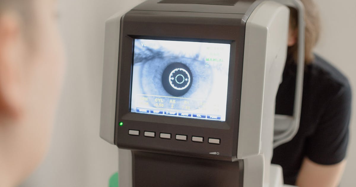
Slit Lamp Examination
The primary diagnostic tool. The ophthalmologist uses a slit lamp (biomicroscope) to examine the lens under high magnification, grading the cataract's density and location. The famous LOCS III grading system is used: nuclear cataract (NO1–NO6), cortical (C1–C5), and posterior subcapsular (P1–P5).
Visual Acuity Testing (Snellen Chart)
Measures how well you see at different distances, expressed as 6/6 (normal vision), 6/9, 6/18, 6/60, and so on. Cataract surgery is generally recommended when visual acuity drops to 6/18 or worse in the better eye, though this is a guideline — quality of life assessment matters more.
Intraocular Pressure (IOP) — Tonometry
Measures pressure inside the eye to rule out glaucoma, which can co-exist with cataract.
Retinal Examination (Fundoscopy/Dilated Eye Exam)
Crucially important before surgery. If the retina is damaged (from diabetic retinopathy, macular degeneration, or retinal detachment), surgery will not restore good vision even after cataract removal. This sets realistic expectations and can change the surgical approach.
Biometry (IOL Power Calculation) — The Key Pre-operative Test
Before surgery, the eye doctor measures the axial length of the eye (using A-scan ultrasound or optical biometry like the IOLMaster) and the corneal curvature (keratometry). These measurements calculate the exact power of the intraocular lens (IOL) that will be implanted — customised for your eye to give you the best possible vision post-surgery.
Modern biometry with optical coherence-based devices (like the IOLMaster or Lenstar) is more accurate than older A-scan ultrasound biometry and is available at most good eye hospitals in Indian cities.
Cost of pre-operative assessment: ₹500–3,000 at private eye hospitals; usually included in the surgery package at larger chains like Sankara Nethralaya, L V Prasad Eye Institute, Aravind Eye Hospital, or Dr. Shroff's Charity Eye Hospital.
Cataract Surgery in India: Techniques Explained
There are two main surgical techniques used in India:
Phacoemulsification (Phaco) — The Gold Standard
Phacoemulsification (commonly called "phaco") is the modern, minimally invasive technique used at all reputable eye hospitals in India. It accounts for 76–88% of cataract surgeries at well-equipped centres.
How it works:
- A tiny incision (2–2.8 mm) is made at the edge of the cornea
- A circular opening is made in the front membrane of the lens capsule
- An ultrasonic probe is inserted; it vibrates at 40,000 cycles per second, emulsifying (liquefying) the hard lens nucleus into small pieces
- The emulsified lens material is aspirated (sucked out)
- A foldable intraocular lens (IOL) is folded, inserted through the tiny incision, and unfolds inside the eye in the correct position
- The incision is self-sealing — usually no stitches required
Recovery: Most patients see significantly better within 24–48 hours. Return to work in 1–3 days for desk work. Avoid strenuous activity, water entering the eye, and heavy lifting for 2–4 weeks.
Manual Small Incision Cataract Surgery (MSICS)
MSICS uses a slightly larger incision (5–7 mm) and removes the lens nucleus in one piece without phacoemulsification. It is:
- Equally effective in skilled hands, with similar visual outcomes to phaco
- Significantly cheaper — making it the technique of choice in government hospitals and high-volume rural camps
- Well-suited for mature, dense cataracts where phaco may be difficult
- Requires a suture in some cases; slightly longer healing time (1–2 weeks)
India's achievement in cataract surgery — bringing high-volume, high-quality surgery to rural areas — is in large part thanks to MSICS, pioneered by surgeons at Aravind Eye Hospital and the now-defunct CBM.
Types of Intraocular Lenses (IOLs): What to Choose
The IOL is the artificial lens implanted to replace the cloudy natural lens. The choice of IOL significantly affects your post-operative vision — and cost.
| IOL Type | What It Corrects | Pros | Cons | Cost Range |
|---|---|---|---|---|
| Monofocal IOL | Either distance OR near vision (you choose) | Best clarity, lowest cost | Still need glasses for the other distance | ₹3,000–8,000/eye |
| Toric IOL | Distance vision + astigmatism | Corrects pre-existing astigmatism | More expensive; positioning critical | ₹15,000–40,000/eye |
| Multifocal/EDOF IOL | Both distance AND near vision (spectacle independence) | Reduces dependence on glasses | Glare/halos possible; not ideal for all patients | ₹25,000–70,000/eye |
| Trifocal IOL | Distance, intermediate, and near | Best spectacle independence | Highest cost; night vision adjustment period | ₹40,000–80,000/eye |
Which IOL is right for you? This depends on your lifestyle, occupation, budget, and the health of your cornea and retina. Discuss with your surgeon — a monofocal IOL gives excellent vision with glasses, while premium IOLs like trifocals are worth considering for active, younger patients who want to be glasses-free.
Cataract Surgery Costs in India
Cost varies enormously based on technique, IOL type, hospital, and city:
| Type of Surgery + IOL | Cost Per Eye | Typical Setting |
|---|---|---|
| Government / Free camp | Free–₹5,000 | Government hospitals, NGO camps (MSICS) |
| Basic Phaco + Monofocal IOL | ₹15,000–30,000 | Mid-tier private hospital |
| Phaco + Toric IOL | ₹25,000–50,000 | Good private eye hospital |
| Phaco + Multifocal/EDOF IOL | ₹40,000–80,000 | Premium eye hospital |
| Femtosecond Laser-assisted (FLACS) | ₹60,000–1,20,000 | Top-tier centres |
Insurance coverage: Cataract surgery is covered under most Indian health insurance policies, including the ₹5 lakh Ayushman Bharat PM-JAY scheme for eligible beneficiaries. CGHS, ESI, and state government health schemes also cover cataract surgery. Always check with your insurer before scheduling.
Free government programmes: Under the National Programme for Control of Blindness and Visual Impairment (NPCB&VI), India set a target of 75 lakh cataract surgeries in FY 2022-23 — and exceeded it with 83.5 lakh surgeries performed. Free cataract surgery camps (Netra Jyoti Abhiyan) are regularly organised across India. Your local district hospital or community health centre can provide information.
Reputed affordable eye hospital chains across India include Aravind Eye Hospitals (Tamil Nadu, Andhra Pradesh), Sankara Nethralaya (Chennai), L V Prasad Eye Institute (Hyderabad), Dr. Shroff's Charity Eye Hospital (Delhi), and Vasan Eye Care (pan-India). These offer subsidised or free surgery based on economic status.
What to Expect: Before, During and After Surgery
Before Surgery
- A pre-operative evaluation is done (slit lamp, IOL biometry, retinal check)
- Blood tests (CBC, blood sugar, ECG) are requested by the anaesthesiologist — surgery is often done under local anaesthesia (eye drops or a small injection around the eye), but your systemic health is still assessed
- Stop blood thinners if prescribed (consult your surgeon and physician)
- Do not eat or drink for 4–6 hours before surgery (for planned sedation)
- Arrange for someone to accompany you — you will not be able to drive home
During Surgery
- Duration: 15–25 minutes for phacoemulsification in uncomplicated cases
- Done under local anaesthesia (topical eye drops + optional IV sedation) — you are awake but relaxed and feel no pain
- A transparent plastic eye shield (not a bulky eye patch) is placed over the eye post-surgery
After Surgery: Recovery Tips
Week 1 (Critical Period):
- Use antibiotic and anti-inflammatory eye drops as prescribed — typically 4–6 times daily; do NOT miss doses
- Wear the protective eye shield while sleeping for 2 weeks
- No water entering the eye — use a towel to wipe the face; no swimming or face-wash directly on the eye for 4 weeks
- Avoid rubbing or pressing on the eye
- No heavy lifting or strenuous exercise for 2 weeks
- You may watch TV, use mobile phones, and read within 48 hours
Week 2–4:
- Vision continues to stabilise; some fluctuation is normal
- Colours may seem brighter and bluer than before surgery (the natural yellowed lens was filtering blue light)
- Mild glare and halos around lights are common initially, especially with multifocal IOLs — usually improves over 2–3 months
Final glasses prescription: Do NOT get new glasses for 6–8 weeks after surgery, as the eye's refraction takes time to fully stabilise.
When to call your doctor urgently:
- Sudden severe pain in the operated eye
- Significant new loss of vision (not just minor fluctuation)
- Increasing redness, discharge, or swelling
- Floating spots, flashes of light, or a shadow/curtain in vision (signs of retinal detachment — rare but requires emergency care)
Diabetics and Cataract Surgery: What to Know
Diabetics in India are at higher risk of cataracts — often developing them 10–15 years earlier than non-diabetics, and the PSC (posterior subcapsular) type is more common. Additionally, diabetics require special care around surgery:
- HbA1c must ideally be below 8% before surgery to reduce infection risk and improve wound healing; discuss with both your ophthalmologist and diabetologist
- Pre-operative fundus examination is essential — if proliferative diabetic retinopathy or macular oedema is present, this needs treatment before or along with cataract surgery, or the visual outcomes will be poor
- Blood sugar control must be optimal in the post-operative period
Tracking Your Eye Health Records on MedicalVault
Cataract surgery generates multiple important documents: pre-operative biometry reports, IOL specifications, surgical records, and post-operative follow-up prescriptions. These are valuable for:
- Future reference if you need additional eye procedures
- Your family members' records (cataracts have a genetic component — if a parent needed surgery at 60, their children should be screened from 50 onwards)
- Insurance claims — having your surgical records organised speeds up reimbursement
Upload your eye examination reports and surgical documents to MedicalVault so they are always accessible — whether you're at a follow-up appointment, travelling, or managing your parents' healthcare from another city. For diabetics tracking multiple conditions, MedicalVault's trend analysis helps you monitor HbA1c, lipid profile, and kidney function alongside your eye health records — all the parameters that affect cataract risk and surgical outcomes.
If you are managing an elderly parent's cataract workup, use MedicalVault's family sharing feature to keep all their records — eye tests, blood reports, and surgical documents — in one organised place.
Key Takeaways
- Cataract is the leading cause of blindness in India (62.6% of all blindness), but it is completely curable with routine surgery.
- India performs over 83 lakh cataract surgeries annually; free surgery is available under government schemes including Ayushman Bharat PM-JAY and the NPCB&VI Netra Jyoti Abhiyan.
- Do not wait for the cataract to become "ripe" — modern surgery is safe and effective at any stage, and early surgery gives better outcomes.
- Phacoemulsification (phaco) is the gold standard, using ultrasound to remove the lens through a tiny incision with rapid recovery; MSICS is equally effective at lower cost in government settings.
- IOL choice matters: monofocal lenses give excellent vision with glasses; toric, multifocal, and trifocal IOLs can reduce glasses dependence but cost more.
- Diabetics need careful pre-operative assessment of blood sugar control and retinal health before cataract surgery.
- Post-operative eye drops must be used diligently for 4–6 weeks; the risk of complications is low in skilled hands.
- Organise your pre- and post-operative eye records on MedicalVault for easy access during follow-ups, insurance claims, and family health management.