The surgeon said the lump "looked harmless", but sent a piece of it to the lab anyway. Now, five anxious days later, the report is on your phone as a PDF: two pages of words like "fibroadenoma", "no atypia", "margins free" or, worse, "infiltrating ductal carcinoma, grade 2, LVI present". Your appointment is not until next week, and the internet is no help at all.
A biopsy report, also called a histopathology report or HPE report, is often the single most important document in a medical journey. It is the test that confirms or rules out cancer, decides whether surgery is needed, and guides chemotherapy, radiation and targeted drugs. India sees an estimated 15 lakh new cancer cases a year, according to ICMR data, and many more biopsies are done for non-cancerous conditions such as skin diseases, kidney problems, liver disease and infections. This guide explains the types of biopsy, how a histopathology report is structured, what common terms mean, how to understand grade, stage and margins, and why you should keep your slides and blocks safe.
What Is a Biopsy and Histopathology?
A biopsy is the removal of a small piece of tissue from the body so that it can be examined under a microscope. Histopathology is the study of that tissue by a pathologist, a doctor trained to diagnose disease from cells and tissues.
How the tissue becomes a report
- Collection: The doctor removes tissue and places it in a preservative, usually 10% formalin.
- Grossing: In the lab, the pathologist examines the specimen with the naked eye, measures it, notes its appearance, and cuts representative pieces.
- Processing: The tissue is dehydrated and embedded in wax to make a paraffin block.
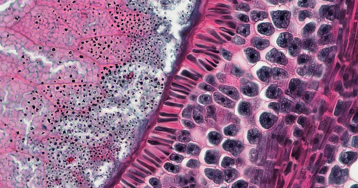
- Sectioning and staining: Very thin slices are cut from the block, placed on glass slides, and stained, usually with haematoxylin and eosin (H&E), which colours cells pink and purple.
- Microscopy: The pathologist examines the slides and writes the report.
- Special tests: If needed, extra stains, immunohistochemistry (IHC) or molecular tests are done on the same block.
Common types of biopsy
| Type | How it is done | Typical uses |
|---|---|---|
| FNAC (fine needle aspiration cytology) | A thin needle draws out cells | Thyroid nodules, lymph nodes, breast lumps; quick and cheap, but gives cells, not tissue structure |
| Core needle biopsy | A thicker needle removes a thin cylinder of tissue, often under ultrasound or CT guidance | Breast, liver, kidney, prostate, lung |
| Incisional biopsy | Part of a lump is removed | Large lesions, oral and skin lesions |
| Excisional biopsy | The whole lump is removed | Small lumps, moles, lymph nodes |
| Punch biopsy | A small circular blade removes a skin sample | Skin rashes, vitiligo, suspected skin cancer |
| Endoscopic biopsy | Taken during endoscopy or colonoscopy | Stomach, food pipe, colon, airway |
| Surgical resection specimen | The entire organ or tumour removed during surgery | Final staging after cancer surgery |
FNAC is cytology, not histopathology. It looks at individual cells, which is useful for screening, but it cannot always show whether a cancer is invasive. A doctor may recommend a core or excision biopsy after an FNAC report says "suspicious" or "inconclusive".
Frozen section
During some cancer surgeries, the surgeon sends tissue for a frozen section, a rapid report within about 20 to 30 minutes, to decide whether to remove more tissue while the patient is still on the table. The final report, based on proper paraffin processing, comes later and can occasionally differ.
How a Histopathology Report Is Structured
Most Indian lab reports follow a similar layout.
| Section | What it contains | What to look for |
|---|---|---|
| Patient and specimen details | Name, age, sex, hospital ID, biopsy or lab number, date of collection and reporting | Check that your name and the body site are correct |
| Clinical details | The doctor's notes, such as "breast lump, 3 cm, left side" | Helps the pathologist interpret findings |
| Specimen | What was sent: for example, "core biopsy, left breast" | Matches what you were told |
| Gross description | Size, weight, colour and appearance of the tissue, and how it was cut | Usually technical; tumour size is often mentioned here |
| Microscopic description | What the pathologist saw under the microscope | Cell patterns, abnormal features |
| Diagnosis / Impression | The final conclusion | The most important line |
| Comment / Note | Recommendations, such as IHC, correlation with scans, or a repeat biopsy | Read this carefully |
| Synoptic report | A checklist format used for cancer, listing grade, margins, lymph nodes and stage | Key information for cancer treatment |
The report should be signed by a qualified pathologist with their registration number. If the lab is accredited, the NABL details will be on the report.
Common Terms in a Biopsy Report
Benign or malignant?
| Term | Meaning |
|---|---|
| Benign | Not cancer; does not spread to other parts of the body |
| Malignant | Cancer; can invade nearby tissues and spread |
| Carcinoma | Cancer starting in lining cells (skin, breast, lung, colon, mouth) |
| Sarcoma | Cancer of bone, muscle, fat or connective tissue |
| Lymphoma | Cancer of lymph glands |
| Inflammation (acute or chronic) | The body's response to infection or injury, not cancer |
| Granuloma / granulomatous inflammation | A specific pattern of inflammation; in India, often due to tuberculosis, especially if "caseating necrosis" is mentioned |
| Hyperplasia | Increased number of normal-looking cells; usually benign |
| Metaplasia | One type of normal cell replaced by another, often due to irritation (as in Barrett's oesophagus) |
| Dysplasia | Abnormal-looking cells that are not yet cancer; graded as mild, moderate or severe (high grade) |
| Atypia | Cells that look unusual; may need follow-up or more tissue |
| Carcinoma in situ | Cancer cells confined to the surface layer without invasion; highly curable |
| Invasive / infiltrating | Cancer that has grown beyond the surface layer |
| Necrosis | Dead tissue |
| Metastatic | Cancer that has spread from somewhere else |
| Negative for malignancy | No cancer seen in the sample |
Suspicious, inconclusive or "not representative"
Sometimes a report says "suspicious for malignancy", "inadequate sample" or "non-representative". This means the pathologist could not reach a firm conclusion, usually because the sample was too small or did not include the abnormal area. It does not mean you have cancer, nor does it rule it out. Your doctor may recommend IHC, a repeat biopsy or a bigger sample.
Understanding Grade, Stage and Margins
If a biopsy shows cancer, the report usually describes several features that guide treatment.
Grade and differentiation
Grade describes how abnormal the cancer cells look and how quickly they are likely to grow.
| Grade | Differentiation | Meaning |
|---|---|---|
| Grade 1 | Well differentiated | Cells look close to normal; usually slower growing |
| Grade 2 | Moderately differentiated | Intermediate |
| Grade 3 | Poorly differentiated | Cells look very abnormal; usually faster growing |
| Undifferentiated | Anaplastic | Very aggressive appearance |
Some cancers have their own grading systems, such as the Gleason score or Grade Group for prostate cancer and the Nottingham grade for breast cancer.
Stage: the pTNM system
Stage describes how far the cancer has spread. After surgery, the pathologist reports a pathological stage, written as pTNM:
- pT: size and extent of the main tumour (pT1 to pT4)
- pN: spread to nearby lymph nodes (pN0 means no spread)
- M: spread to distant organs, usually decided by scans rather than the biopsy
A prefix "y" (as in ypT2) means the stage was measured after chemotherapy or radiation given before surgery. Your oncologist combines this information to assign an overall stage from I to IV.
Margins
When a tumour is removed, the pathologist checks whether the edges of the specimen are free of cancer.
- Negative, clear or free margins: No cancer at the edge; the tumour was likely fully removed.
- Close margin: Cancer is very near the edge (the report may give the distance in mm).
- Positive or involved margin: Cancer cells reach the edge; some may remain. More surgery or radiation may be advised.
Other features that affect treatment
- Lymphovascular invasion (LVI): Cancer cells seen in small blood or lymph vessels, which raises the risk of spread.
- Perineural invasion (PNI): Cancer growing along nerves, common in head and neck cancers.
- Lymph nodes: Reported as, for example, "3/18 nodes positive", meaning cancer was found in 3 of 18 nodes examined.
- Depth of invasion (DOI): Important in oral cancer, which is very common in India because of tobacco and gutka use. See our oral cancer guide.
- Tumour size: Usually given in centimetres.
IHC and Molecular Tests
Immunohistochemistry (IHC) uses special antibodies to identify proteins in the tissue. It helps confirm the type of cancer, find where a cancer originated, and predict response to treatment.
| Test | Used in | Why it matters |
|---|---|---|
| ER, PR (oestrogen and progesterone receptors) | Breast cancer | Positive tumours respond to hormone therapy |
| HER2 | Breast and stomach cancer | Positive tumours may benefit from targeted drugs such as trastuzumab |
| Ki-67 | Breast and other cancers | Shows how fast cells are dividing |
| PD-L1 | Lung, head and neck and other cancers | Helps decide on immunotherapy |
| CD markers (CD20, CD3 and others) | Lymphomas | Determine lymphoma type and treatment |
| EGFR, ALK, ROS1 (molecular) | Lung cancer | Identify patients for targeted tablets |
| KRAS, NRAS, BRAF, MSI | Colorectal cancer | Guide targeted therapy and immunotherapy |
Molecular tests usually need the original paraffin block and may be done at a different lab. Our breast cancer guide and lung cancer guide explain how these results shape treatment. Blood tumour markers are different from tissue tests; see our tumour marker test guide.
Time and cost in India
| Test | Typical time | Approximate cost |
|---|---|---|
| Small biopsy (skin, endoscopic, core) | 3 to 7 working days | ₹1,000 to ₹3,000 |
| Large surgical specimen | 5 to 10 working days | ₹3,000 to ₹10,000 or more |
| IHC (per marker or panel) | Adds 2 to 5 days | ₹1,500 to ₹3,000 per marker; panels cost more |
| Molecular tests | 1 to 3 weeks | Several thousand to tens of thousands of rupees |
| Second opinion review of slides and blocks | 4 to 7 working days | Roughly ₹1,500 to ₹10,000, depending on IHC |
Costs are much lower at government hospitals and regional cancer centres, and cancer diagnosis and treatment are covered under Ayushman Bharat PM-JAY for eligible families.
Slides, Blocks and Second Opinions
Why your slides and blocks matter
Your paraffin blocks and slides are not just lab waste. They contain your tissue, and they may be needed:
- For a second opinion at another hospital
- For IHC or molecular tests ordered later
- When a cancer recurs years later, so it can be compared with the original
- When you change hospitals or cities for treatment
Labs keep blocks and slides for several years. You can request them, usually on loan, by submitting a written application and paying a small fee. Read our guide on your right to medical records for how to ask for copies.
When to seek a second opinion
Pathology diagnoses, especially for cancer, lymphoma and rare tumours, can be difficult. A second opinion from an experienced cancer pathologist is reasonable when:
- The diagnosis will lead to major surgery, chemotherapy or radiation
- The report says "suspicious", "atypical" or "cannot rule out"
- The cancer type is rare or unusual
- The report seems to conflict with scans or the clinical picture
Major cancer centres such as Tata Memorial Hospital offer pathology review services; you send the slides, blocks and the original report. Our online second opinion guide explains how to arrange this.
Questions to ask your doctor
- Is this benign or malignant? Is this the final diagnosis?
- What is the grade and, if applicable, the stage?
- Were the margins clear?
- Are further tests, such as IHC or molecular tests, needed?
- Do I need a repeat biopsy or more surgery?
- How will this report change my treatment?
Keeping Biopsy Reports and Records Safe
A biopsy report is a document you may need for the rest of your life. Cancer survivors are asked for their original histopathology at every follow-up, when they consult new specialists, buy insurance or apply for disability benefits. Yet the report is often a single sheet in a hospital file, or a PDF lost in a WhatsApp chat.
When you upload your reports to MedicalVault, your biopsy report, scans and blood tests are organised in one place, so the original histopathology is always at hand at every appointment. MedicalVault's trend analysis lets you follow blood tests such as haemoglobin and tumour markers during and after treatment. With the family sharing feature, the family member who handles your appointments can see the same reports, which helps during the stressful weeks of diagnosis and treatment.
Key Takeaways
- A biopsy report (histopathology or HPE report) is the definitive test for diagnosing cancer and many other conditions.
- FNAC looks at cells (cytology), while core, incisional and excisional biopsies provide tissue (histopathology), which gives more information.
- Focus on the diagnosis or impression line, the comment, and, for cancer, the synoptic report with grade, margins, lymph nodes and pTNM stage.
- Dysplasia and carcinoma in situ are early changes, while invasive carcinoma has grown beyond the surface; granulomatous inflammation in India often points to TB.
- Clear margins, a lower grade and no lymph node or vessel involvement generally mean a better outlook, but your oncologist interprets the full picture.
- IHC (ER, PR, HER2, Ki-67, PD-L1) and molecular tests guide targeted treatment and need the original paraffin block.
- Keep your slides, blocks and biopsy report safe, consider a second opinion for major decisions, and store every report in MedicalVault.